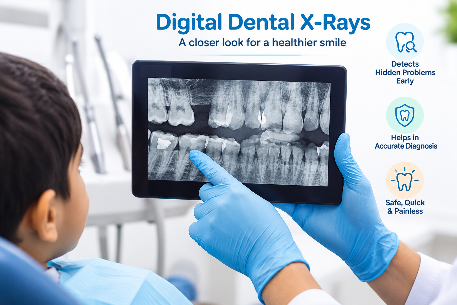
Why Dental X-Rays Are Essential for Children
A visual examination alone reveals only what can be seen on the surface of teeth and gums. Up to 40% of tooth decay occurs between teeth — in contact areas that are completely invisible without radiographs. Dental X-rays also show:
- Cavities forming between teeth before they are visible or symptomatic
- The position and development of unerupted permanent teeth
- Bone levels around teeth — critical for assessing infection spread
- Root abnormalities, cysts, or extra (supernumerary) teeth
- Spacing available for erupting permanent teeth — guiding orthodontic planning
Skipping radiographs means treating only what is visible and missing early decay that could be addressed with a simple filling before it reaches the nerve.
Digital vs Film X-Rays
Dr. Mittal's clinics use exclusively digital radiography. Digital sensors require significantly less radiation to produce a diagnostic image compared to conventional film, and the images are instantly available on screen — no developing chemicals, no waiting, and instant enlargement for detailed review with the parent present.
Digital images can also be stored indefinitely and compared across visits to track changes — showing whether a cavity is stable or progressing year-over-year.
Types of X-Rays Used
- Bitewing radiographs — show the crowns of upper and lower back teeth simultaneously; detect interproximal (between-teeth) decay and bone levels. Most commonly taken.
- Periapical radiographs — show the entire tooth from crown to root tip; used when assessing infection, abscess, or root damage.
- OPG (Orthopantomogram) — a panoramic view of all teeth, jaw, and developing permanent teeth; taken for orthodontic assessment or when a full mouth overview is needed.
How Often Are X-Rays Taken
Radiographs are taken only when clinically indicated — not automatically at every visit. The frequency depends on the child's cavity risk:
- Low risk — bitewing X-rays every 12–18 months once baby molars are in contact
- Moderate risk — every 6–12 months
- High risk — every 6 months or as clinically needed
Frequently Asked Questions
Yes. Digital dental radiographs produce approximately 90% less radiation than conventional film X-rays. A full set of four bitewing radiographs produces less radiation than a 2-hour flight. A lead apron and thyroid collar are used at every exposure. X-rays are taken only when clinically necessary — not at every visit. The benefit of detecting early decay far outweighs the minimal radiation risk.
Dental X-rays are taken from the age when the baby molars first come into contact with each other — typically around age 2–3 — as this is when interproximal decay between back teeth becomes possible. In practice, the clinical need drives the timing: if a young child has visible cavities or pain, a periapical X-ray may be taken even before age 3. Cooperation is often the limiting factor in very young children.